Home » Without Label » Back Of Neck Anatomy - Labeled Anatomy Chart Of Neck And Back Muscles On White Background Stock Photo Download Image Now Istock - It is made up of bones, discs, muscles, ligaments, nerves and tendons.
Back Of Neck Anatomy - Labeled Anatomy Chart Of Neck And Back Muscles On White Background Stock Photo Download Image Now Istock - It is made up of bones, discs, muscles, ligaments, nerves and tendons.
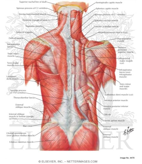
Back Of Neck Anatomy - Labeled Anatomy Chart Of Neck And Back Muscles On White Background Stock Photo Download Image Now Istock - It is made up of bones, discs, muscles, ligaments, nerves and tendons.. The neck is one of the most complex and intricate structures in our body and includes the spinal cord, which sends messages from the brain to the rest of the body. The neck also protects the nerves and the spinal cord as well to help maintain a healthy neck anatomy area diagram body maps. The inner portions of the tooth consist of the dentin, a bonelike tissue, and the pulp. The back anatomy includes the latissimus dorsi, trapezius, erector spinae, rhomboid, and the teres major. The motion of the muscles of the neck are divided into four.
The cervical spine supports the weight and movement of your head and protects the nerves exiting your brain. Bones of the neck picture. The motion of the muscles of the neck are divided into four. Below the neck, holding the tooth into the bone, is the root of the tooth. An area called the occiput.
Muscles Of The Neck And Torso Classic Human Anatomy In Motion The Artist S Guide To The Dynamics Of Figure Drawing from doctorlib.info They collect lymph from the posterior neck, upper ear and the back of the external auditory meatus (the ear canal). The larynx is located where the pharynx, the back of the mouth and nasal cavity, divides into the trachea (the tube that carries air to the lungs) and the esophagus (the tube that carries food to. They move the head in every direction, pulling the skull and jaw towards the shoulders, spine, and scapula. It consists of seven vertebrae. Neck muscles can be strained from poor posture — whether it's leaning over your computer or hunching over your workbench. Instant anatomy is a specialised web site for you to learn all about human anatomy of the body with diagrams, podcasts and revision questions. Top head neck anatomy flashcards ranked by quality. Bones of the neck picture.
The hard white exterior covering of the tooth is the enamel.
The cervical spine, your neck, is a complex structure making up the first region of the spinal column starting immediately below the skull and ending at the first thoracic vertebra. Causes of neck pain and how to manage the pain in basic terms, the neck (cervical spine) joins the shoulders and chest to the head. They collect lymph from the posterior neck, upper ear and the back of the external auditory meatus (the ear canal). It runs down the back part of the neck, and opens into the external jugular vein just below the middle of its. The cervical spine protects the nerves connecting to the brain, allowing the head to move freely while supporting its weight. Jugularis posterior) begins in the occipital region and returns the blood from the skin and superficial muscles in the upper and back part of the neck, lying between the splenius and trapezius. The larynx is located where the pharynx, the back of the mouth and nasal cavity, divides into the trachea (the tube that carries air to the lungs) and the esophagus (the tube that carries food to. Back pain is common and might be caused by a problem with a muscle. The majority of these nerves control the functions of the upper extremities and allow you to feel your arms, shoulder, and back of your head. Muscle head anatomy vocal organ diagram female neck anatomy neck wireframe head neck human anatomy head artery anatomy face pharynx vector neck degree head anatomy 3d. An area called the occiput. The suboccipital muscles act to rotate the head and extend the neck.rectus capitis posterior major and rectus capitis posterior minor attach the inferior nuchal line of the occiput to the c2 and c1 vertebrae respectively.obliquus capitis superior also extends from the occiput to c1 while obliquus capitis inferior originates from c2 and. The posterior external jugular vein (v.
It runs from the neck to the upper back. Osteoarthritis also is a common cause of neck pain. The neck is connected to the upper back through a series of seven vertebral segments. The cervical spine the neck and upper back composed of the seven vertebrae closest to the skull. Bones of the neck picture.
Upper Cervical Spine Disorders Anatomy Of The Head And Upper Neck from www.spineuniverse.com Labeled anatomy chart of neck and back muscles on black background labeled human anatomy diagram of man's neck and back muscles from a posterior view on a black background. Rarely, neck pain can be a symptom of a more serious problem. Bones of the neck picture. Think of it like a jigsaw puzzle, all the pieces fit in together and are required to get the full picture as to how it works. Choose from 500 different sets of flashcards about neck anatomy back neck upper on quizlet. The anatomy of your back muscles can be complex. Each nerve provides sensation to a specific area of the body called a dermatome. The anterior, and the posterior, triangles of the neck.
It is made up of bones, discs, muscles, ligaments, nerves and tendons.
Neck muscles can be strained from poor posture — whether it's leaning over your computer or hunching over your workbench. Jugularis posterior) begins in the occipital region and returns the blood from the skin and superficial muscles in the upper and back part of the neck, lying between the splenius and trapezius. As the tooth tapers below the gumline, the neck is formed. The majority of these nerves control the functions of the upper extremities and allow you to feel your arms, shoulder, and back of your head. The suboccipital muscles act to rotate the head and extend the neck.rectus capitis posterior major and rectus capitis posterior minor attach the inferior nuchal line of the occiput to the c2 and c1 vertebrae respectively.obliquus capitis superior also extends from the occiput to c1 while obliquus capitis inferior originates from c2 and. It consists of two major parts: The muscles of the neck are present in four main groups. It consists of seven vertebrae. The neck is connected to the upper back through a series of seven vertebral segments. Neck anatomy explained the neck begins at the base of the skull and connects to the thoracic spine the upper back. Labeled anatomy chart of neck and back muscles on black background labeled human anatomy diagram of man's neck and back muscles from a posterior view on a black background. Rarely, neck pain can be a symptom of a more serious problem. They start at the top of the neck and go down to the tailbone.
Anatomy of back of human neck, anatomy of the back and neck, anatomy of the back of the neck, anatomy of the back of the neck muscles, anatomy of the back of your. Neck anatomy explained the neck begins at the base of the skull and connects to the thoracic spine the upper back. Neck anatomy explained the neck begins at the base of the skull and connects to the thoracic spine (the upper back). See spinal cord anatomy in the neck according to the centers for disease control and prevention (cdc), a fever, headache, and stiff neck (inability to flex the neck forward, also called nuchal rigidity) are typically early symptoms of bacterial meningitis.foot|1] when any two of these symptoms are present together, they should be immediately. Osteoarthritis also is a common cause of neck pain.
Atlas Of Human Anatomy 3e from www.netterimages.com Cervical spine anatomy video the cervical spine has 7 stacked bones called vertebrae, labeled c1 through c7. The anterior, and the posterior, triangles of the neck. It runs from the neck to the upper back. See spinal cord anatomy in the neck according to the centers for disease control and prevention (cdc), a fever, headache, and stiff neck (inability to flex the neck forward, also called nuchal rigidity) are typically early symptoms of bacterial meningitis.foot|1] when any two of these symptoms are present together, they should be immediately. The cervical spine, your neck, is a complex structure making up the first region of the spinal column starting immediately below the skull and ending at the first thoracic vertebra. Labeled anatomy chart of neck and back muscles on black background labeled human anatomy diagram of man's neck and back muscles from a posterior view on a black background. The inner portions of the tooth consist of the dentin, a bonelike tissue, and the pulp. Top head neck anatomy flashcards ranked by quality.
Each nerve provides sensation to a specific area of the body called a dermatome.
Bones of the neck picture. See spinal cord anatomy in the neck according to the centers for disease control and prevention (cdc), a fever, headache, and stiff neck (inability to flex the neck forward, also called nuchal rigidity) are typically early symptoms of bacterial meningitis.foot|1] when any two of these symptoms are present together, they should be immediately. In the neck are five groups of lymph nodes that drain lymph from the tissues of the neck: The top of the cervical spine connects to the skull, and the bottom connects to the upper back at about shoulder level. The occipital bone surrounds a large opening known as the foramen magnum. It runs from the neck to the upper back. Top head neck anatomy flashcards ranked by quality. In neck, groin, armpits & throat. Neck anatomy explained the neck begins at the base of the skull and connects to the thoracic spine (the upper back). The neck is connected to the upper back through a series of seven vertebral segments. The hard white exterior covering of the tooth is the enamel. The occipital bone is a bone that covers the back of your head; It is made up of bones, discs, muscles, ligaments, nerves and tendons.